About HKJR
Hong Kong Journal of Radiology (HKJR) is the official peer-reviewed academic journal of the Hong Kong College of Radiologists. HKJR is published quarterly by Hong Kong Academy of Medicine Press. HKJR is a continuation of the Journal of the Hong Kong College of Radiologists.
HKJR publishes papers on all aspects of diagnostic imaging, clinical oncology, and nuclear medicine, including original research articles, review articles, perspectives, pictorial essays, case reports, brief communications, editorials, and letters to the Editor. Papers on radiological protection, quality assurance, audit in radiology, and matters related to radiological training or education are also included.
The 2023 Journal Impact Factor for the HKJR is 0.2 (Clarivate, 2024).
FREE full text of ALL issues is available.
Additional materials may be made free at the Editorial Board's discretion.
Online First articles
Online First articles are released before they are included in a journal issue. These articles are fully citable and come with a DOI, enabling the most recent research to be accessed promptly.
Current Issue
Volume 28 Number 1, March 2025
![]() FULL TABLE OF CONTENTS Download the full issue
FULL TABLE OF CONTENTS Download the full issue
Highlights of this issue
About the Cover Images
 |
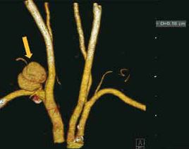 |
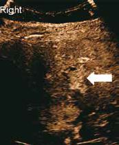
| In the article “Artificial Intelligence for Contouring and Treatment Planning in Locoregional Radiotherapy Including Internal Mammary Nodal Irradiation for Breast Cancer”. Images from the same patient at two different levels showing the dose distribution achieved by volumetric modulated arc therapy created by machine learning. | In the article “Magnetic Resonance Imaging of Brachial Plexus Pathologies: A Pictorial Essay”. Coronal fat-suppressed T2-weighted image showing oedema of left brachial plexus in a patient with acute left brachial neuritis (arrows). |